Oxygen Transport by Hemoglobin and Myoglobin

Requires a Wolfram Notebook System
Interact on desktop, mobile and cloud with the free Wolfram Player or other Wolfram Language products.
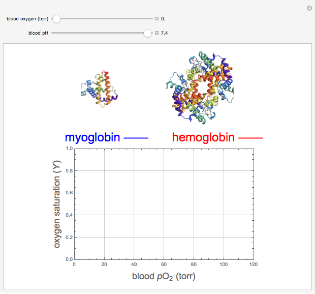
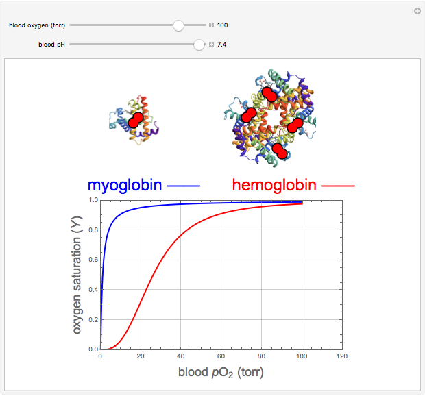
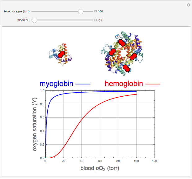
Hemoglobin, the essential component of red blood cells (erythrocytes), transports oxygen ( ) through the bloodstream from the lungs to all the tissues of the body. Hemoglobin also carries carbon dioxide (
 ) back to the lungs to complete the process of respiration. Vertebrate hemoglobin is a nearly spherical protein molecule consisting of an array of four globin polypeptide chains, each containing a heme group, which is the oxygen-binding site. Its molecular weight is approximately 64,500 daltons. The oxygen uptake of hemoglobin exhibits cooperativity, such that each
  successively increases the affinity of the molecule for adding another
  , up to four. The saturation
  is a measure of the fractional occupancy of the oxygen-binding sites. It increases as a sigmoid-shaped function of the partial pressure of
  in its immediate environment. In the alveoli of the lungs,
  is approximately 100 torr.
Contributed by: S. M. Blinder (April 2011)
Open content licensed under CC BY-NC-SA
Snapshots
Details
The binding of oxygen to myoglobin is described by the equilibrium  . The saturation can be defined by
  , where
  is the partial pressure for 50% saturation, about 1 torr for myoglobin. For hemoglobin, the equilibrium takes the form
  ,
,  to
to  . Here
  , where
  to account for the cooperativity of
  binding and
  at pH 7.4 but slightly higher as the pH decreases.
The central part of the heme group has this structure:  . The
. The  complexes with the iron atom.
Reference: L. Stryer, Chap. 7, Molecular Design of Life, New York: W. H. Freeman, 1989.
Permanent Citation
